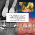

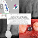
Extraction of a Fractured Upper Left Second Premolar, Conventional Implant Placement, with Bone Grafting and Immediate Loading of a Provisional. Final restoration with digital occlusal analysis using T-Scan. – A Case Report
Dr Fabrizia Luongo, DDS, MS, Periodontist, Rome, Italy
Introduction
The evolution of digital dentistry and the development of a digital workflow has concentrated on digital planning with the use of Cone Beam CT scanning as well as using digital restorative tools to combine DICOM (Digital Imaging Communication in Medicine) and .stl (stereolithography) files to virtually plan, place and restore implants before using this plan to treat patients. The resulting benefits are reduced chair time, high precision and predictable aesthetic results often with immediate fixed provisional restorations available at time of surgery and corresponding high levels of patient satisfaction.
Intra-oral scanning to create digital ‘virtual impressions’ is also becoming more prevalent with the information being stored in the .stl file format. This information can be utilised by appropriate CAD/CAM (computer-aided design and computer aided manufacturing) software to design and manufacture a dental restoration (either by milling or 3D printing).
One area that is sometimes overlooked is the use of digital technology in occlusal analysis and adjustment of the restored dental implant. The following case study examines the occlusal management of a conventionally placed implant.