Catarina G. Rodrigues, DDS, MSc – Manuel D. Marques, DDS
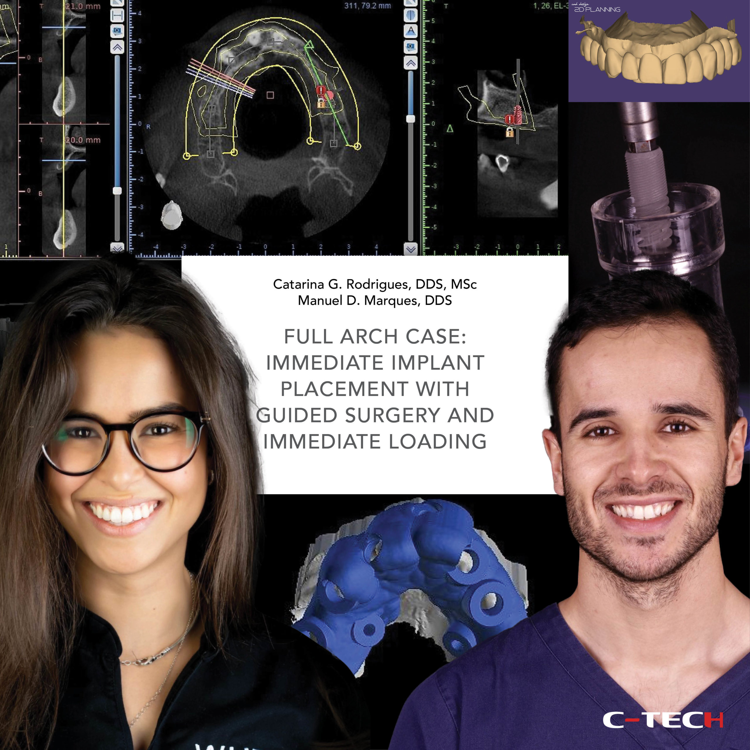
In the present clinical case, the upper left lateral incisor presented with a vertical fracture. Following a proper clinical and radiographic analysis, the tooth was considered hopeless. The treatment plan consisted of the extraction of the lateral incisor and immediate dental implant placement. It is well described in the literature that delayed loading, in contrast with immediate or immediate- delayed loading, can lead to predictable results in all clinical situations.
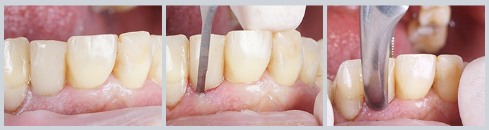
For that reason, it is highly recommended in particular conditions, such as dental implants of reduced dimensions, and compromised host conditions. In this specific case, to reduce risks, a delayed loading protocol was followed. Nonetheless, to preserve the pre-operative soft tissue architecture which was ideal (Fig.1), a customized healing abutment was fabricated. Also, immediate aesthetics was ensured with an adhesive bridge using the patient’s own tooth.
Following local anesthesia, a sharp dissection of the supracrestal fibers with a micro scalpel blade was performed and the tooth was removed carefully with extraction forceps (Fig. 2 and 3).
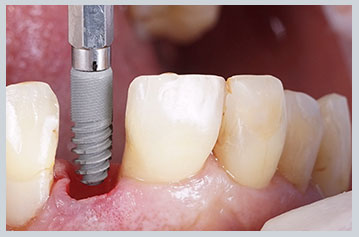
The socket was thoroughly debrided with a surgical excavator and rinsed with saline. The integrity of the buccal wall was verified. A 3.8×11 mm dental implant (C-Tech Implant) was placed at the lateral incisor site (Fig. 4).
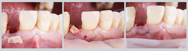
To compensate for the changes in the soft tissue contour that spontaneously occur following a tooth extraction, which can have a negative impact on the health of peri-implant tissues, and compromise the final aesthetic result, a simultaneous soft tissue augmentation, at the time of dental implant placement, using a connective tissue graft was performed (Fig 5-7).
 Then, using a peek provisional abutment (C-Tech Implant) and flowable composite, a customized healing abutment was fabricated to support local anatomy during the osseointegration phase (Fig. 8). With this approach, after osseointegration, we can obtain an anatomic representation that mimics the emergence profile of the natural dentition.
Finally, immediate aesthetics was achieved using the patient’s own tooth. The anatomical crown of the extracted tooth was steam cleaned, treated, and prepared to obtain a facial veneer that was used to fabricate a provisional adhesive bridge (Fig. 9 and 10).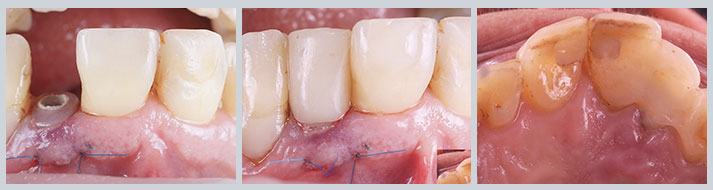 The utilization of the patient׳s own tooth can provide a seamless and comfortable transition from hopeless teeth to provisional restoration.