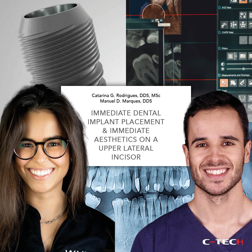
Dr Kevin Briffa B.Ch.D, M.Sc Oral Implantology (Fran.) – Mysmile Dental Care Centre, Malta
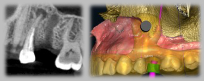
A 44 year old female patient presented to a private dental clinic requesting to replace her missing upper left second premolar. A CBCT radiograph was taken to plan the case. An incidental finding was that there was a periapical lesion associated with the roots of the asymptomatic upper left first premolar. This tooth had a root-canal filling and its roots curved towards the site where the implant would be placed. An intraoral scan was taken and this was superimposed onto the CBCT to plan the surgical guide.
The surgical guide was designed to serve for two purposes:
– for guided surgery implant insertion
– to locate the exact site of the periapical lesion to carry out minimally-invasive surgery when carrying out the apicectomy procedure
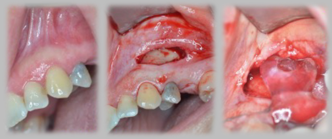
During the surgery appointment, an incision was made in the buccal mucosa at a level slightly crestal to the area of the periapical lesion. A flap was raised and the tooth-supported guide was placed onto the teeth, ensuring that it was fitting perfectly.
Trephine burs were used through the ‘window’ designed in the surgical guide to remove a ‘lid’ of bone overlying the periapical lesion. A significant part of the granuloma came out attached to the bony ‘lid’ when this was removed.
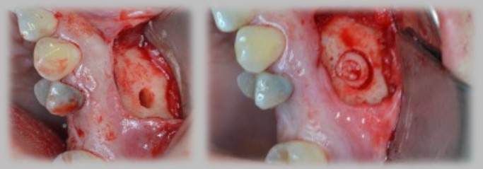
After completion of the apicectomy procedure and proper degranulation of the infected area, the bony ‘lid’ was placed back in its original place.
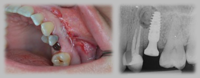
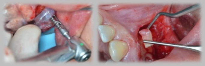
The apicectomy flap was sutured with PTFE sutures. The standard guided surgery routine procedure was subsequently carried out for the placement of a C-Tech EL 4.3x11mm implant to replace the missing upper left second premolar. The post-operative periapical radiograph shows the implant with its healing abutment and the completed apicectomy of the upper left first premolar, with the replaced bony ‘lid’ overlying the surgical site.